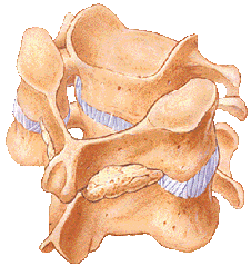
Posterior Cervical Fusion
What is the Rationale Behind Posterior Cervical Fusion?
The posterior cervical fusion is performed through an incision in the back of the neck. A posterior cervical fusion is used to stop the motion between two or more vertebrae. It is commonly used to prevent spinal deformity from developing after a large decompression; to rectify a spinal deformity; or to stabilize the spine after a fracture or dislocation of the cervical spine.
What is the Procedure of Posterior Cervical Fusion?
This surgery is done through the back of the neck. After the surgeon has excised any bone, tumour or disc fragments which may be compressing the spinal cord and/or the exiting nerve roots, screws are then introduced into the outer parts of the vertebrae from behind, and linked by rods which correct spinal deformity or prevent deformity or instability occurring. Bone graft is then placed on the back surface of the problem vertebrae. During the healing process, the vertebrae grow together, creating a solid piece of bone. This is called a posterior instrumented fusion (see next section).

The rationale of using Instrumentation in Posterior Cervical Fusion:
When instrumentation is used to improve the success of a posterior fusion, metal rods or plates are attached to the bone structures in the back of the spine, to stop movement between the verterbrae so the bone can fuse. Stainless steel or titanium cables can also be used. When doctors use this type of instrumentation, a brace may only be needed for a short period of time, or not at all.
Once the bone graft has fused, the metal usually has no further function, and biomechanics talk of the bone graft “unloading the metal construct”. Metal is almost always incapable of causing ongoing symptoms once the instrumented segments have fused, so metal constructs are seldom removed unless they have become infected.
